
Human anatomy and physiology lab manuals are crucial resources, offering detailed guidance for practical learning experiences. These manuals, like those by Marieb & Smith, enhance understanding through focused exercises.
They provide essential instructions and a structured approach to dissecting and observing anatomical structures, ultimately solidifying theoretical knowledge with hands-on application.
Purpose of a Lab Manual
A human anatomy and physiology lab manual serves as a cornerstone for effective learning, bridging the gap between theoretical concepts and practical application. Its primary purpose is to guide students through a series of carefully designed experiments and observations, fostering a deeper comprehension of the human body’s intricate systems.
These manuals, such as the widely used text by Elaine Marieb and Lori Smith, provide detailed, step-by-step instructions for dissections – like those utilizing a fetal pig model – and physiological experiments. They aren’t merely procedural checklists; they encourage critical thinking and data analysis, essential skills for future healthcare professionals.
Furthermore, a lab manual facilitates the development of essential laboratory techniques, ensuring students are proficient in handling equipment and adhering to safety protocols. Access to resources like Nursing Times, with its clinical articles and procedures, complements the manual’s core function, providing real-world context and enhancing the overall learning experience. Ultimately, the manual aims to transform students from passive learners into active investigators.
Importance of Practical Experience
Practical experience is paramount in mastering human anatomy and physiology, and a lab manual facilitates this crucial learning process. While textbooks provide foundational knowledge, hands-on activities solidify understanding in a way that reading alone cannot. Dissections, like those performed on fetal pigs – a common model – allow students to visualize anatomical structures in three dimensions, enhancing spatial reasoning.
Physiological experiments, such as measuring lung capacity or analyzing urine, demonstrate functional relationships and reinforce theoretical concepts. This active engagement promotes deeper retention and critical thinking skills. Resources like peer-reviewed clinical articles, accessible through subscriptions like Nursing Times, demonstrate the real-world relevance of these skills.
Furthermore, practical work develops essential laboratory techniques, data analysis abilities, and observational skills – all vital for success in healthcare careers. The lab manual, therefore, isn’t just a guide; it’s a gateway to becoming a competent and confident practitioner.
Essential Components of a Lab Manual
Effective lab manuals include safety protocols, material lists, anatomical terminology reviews, and detailed dissection/experiment guides, ensuring a structured and secure learning environment.
Safety Guidelines and Protocols
Prioritizing safety is paramount in any anatomy and physiology laboratory setting. A comprehensive lab manual must dedicate a significant section to detailed safety guidelines and protocols. These guidelines should explicitly cover proper handling and disposal of specimens – particularly crucial during dissections, like those utilizing fetal pigs.
Students need clear instructions on the correct use of dissection tools, emphasizing caution to prevent self-injury or damage to equipment. Personal protective equipment (PPE), including gloves, goggles, and lab coats, should be mandatory, with detailed instructions on their proper donning and doffing.
The manual should also outline emergency procedures, including the location of first aid kits, eyewash stations, and emergency contact information; Specific protocols for handling potentially hazardous materials, like preservatives or stains, are essential. A clear statement regarding appropriate lab attire – closed-toe shoes, tied-back hair – is also vital. Finally, a section on proper hygiene practices, such as handwashing, reinforces a safe learning environment.
List of Required Materials & Equipment
A detailed inventory of required materials and equipment is a cornerstone of any effective anatomy and physiology lab manual. This section should provide a comprehensive list, categorized for clarity, ensuring students are fully prepared for each laboratory session. Essential items typically include dissection kits – containing scalpels, forceps, scissors, and probes – alongside dissecting pans and pins.
Microscopes, slides, and staining solutions are crucial for histological studies. Depending on the experiments, the list may also encompass physiological monitoring equipment, such as spirometers for respiratory measurements or sphygmomanometers for blood pressure assessment.
The manual should specify the quantity and type of each item, along with any specific brand recommendations. Furthermore, it’s beneficial to include information on where to procure these materials, whether through the institution’s bookstore or external suppliers. Clear labeling of equipment and proper storage instructions should also be outlined to maintain organization and longevity.
Anatomical Terminology Review
A robust anatomical terminology review is paramount within a human anatomy and physiology lab manual. This section serves as a foundational refresher, ensuring all students possess a common understanding of the language of the body. It should meticulously define directional terms – superior, inferior, anterior, posterior, medial, and lateral – alongside positional and regional designations.
The review must also cover standard anatomical position, planes of the body (sagittal, frontal, transverse), and common prefixes and suffixes used in anatomical nomenclature.
Illustrations and diagrams are invaluable in reinforcing these concepts. Quizzes or self-assessment exercises can further solidify student comprehension. A glossary of key terms, readily accessible throughout the manual, is also highly recommended. Mastery of this terminology is essential for accurate observation, dissection, and communication within the laboratory setting.
Body System Dissections & Observations
This section details dissections of key systems – skeletal, muscular, nervous, and cardiovascular – enabling students to observe anatomical structures firsthand and correlate form with function.
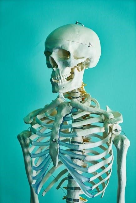
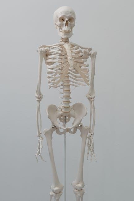
Skeletal System: Bone Identification & Articulations
The skeletal system component of a human anatomy and physiology lab manual focuses on comprehensive bone identification. Students will learn to distinguish various bones – long, short, flat, irregular, and sesamoid – by their unique features and markings.
Practical exercises involve identifying specific bones in skeletal models, diagrams, and potentially, actual skeletal specimens. A key aspect is understanding bone articulations, the points where bones connect.
Students will categorize different types of joints – fibrous, cartilaginous, and synovial – and analyze their range of motion. The manual guides observation of joint structures like ligaments and cartilage, crucial for stability and movement.
Furthermore, the lab will explore bone landmarks, such as processes, foramina, and fossae, relating these features to muscle attachments and nerve pathways. Accurate identification and understanding of articulations are fundamental to grasping skeletal function.
Muscular System: Muscle Identification & Function
The muscular system section within a human anatomy and physiology lab manual emphasizes precise muscle identification. Students will learn to locate and name superficial muscles, utilizing anatomical models and diagrams as primary resources.
Lab exercises concentrate on recognizing muscle origins, insertions, and actions. Understanding these elements is vital for comprehending how muscles produce movement. The manual guides students through palpating (feeling) muscles on themselves and partners, enhancing tactile learning.
A core component involves classifying muscles based on their fiber arrangement – parallel, pennate, and circular – and correlating this structure with functional capabilities. Students will analyze how muscle contractions generate force and facilitate various body movements.
Furthermore, the lab explores muscle groups and their synergistic or antagonistic relationships, demonstrating how muscles work together to achieve coordinated motion. Accurate identification and functional analysis are key to mastering muscular system anatomy.
Nervous System: Brain Dissection & Nerve Pathways
The nervous system component of a lab manual centers on brain dissection and tracing nerve pathways. Students carefully dissect preserved sheep brains, identifying major structures like the cerebrum, cerebellum, and brainstem. Detailed diagrams and illustrations are essential for accurate identification.
Lab exercises focus on recognizing cranial nerves and their associated functions, linking structure to physiological roles. Students will explore the brain’s external features and then proceed to internal dissections, revealing key areas like the thalamus and hypothalamus.
A crucial aspect involves tracing major nerve pathways responsible for sensory and motor functions. This reinforces understanding of how the brain processes information and controls bodily responses. The manual guides students through identifying the spinal cord and its connection to the brain.
Ultimately, this section aims to provide a tangible understanding of the brain’s complex anatomy and the intricate network of nerves that govern the body.
Cardiovascular System: Heart Dissection & Blood Vessels
The cardiovascular system section of the lab manual emphasizes heart dissection and identification of major blood vessels. Typically, preserved sheep hearts are used, allowing students to examine the chambers – atria and ventricles – and associated structures like valves and major arteries/veins.
Detailed illustrations guide students through the dissection process, highlighting the flow of blood through the heart. Lab exercises focus on identifying the aorta, pulmonary artery, superior and inferior vena cava, and pulmonary veins. Students learn to trace the path of blood circulation.
Furthermore, the manual explores the structure of blood vessels – arteries, veins, and capillaries – and their role in transporting blood throughout the body. Microscopic slides may be used to observe vessel wall layers.
This practical experience solidifies understanding of cardiac anatomy and the circulatory system’s function, linking structure to physiological processes like blood pressure and oxygen delivery.
Physiological Experiments & Data Analysis
This section details experiments like lung capacity measurements, enzyme activity, and urine analysis, requiring students to collect data and interpret results for physiological understanding.
Respiratory System: Lung Capacity Measurements
Lung capacity measurements are a fundamental component of a human anatomy and physiology lab, providing practical insight into respiratory function. Lab manuals guide students through techniques to determine vital capacities, including tidal volume, inspiratory reserve volume, expiratory reserve volume, and residual volume.
Typically, students utilize spirometers – devices that measure air movement – to collect data during controlled breathing exercises. The manual will detail proper spirometer usage, calibration procedures, and standardized breathing protocols to ensure accurate results.

Data analysis involves calculating lung capacities and comparing individual results to predicted values based on factors like age, height, and sex. Students learn to interpret variations, potentially identifying factors influencing respiratory efficiency. Furthermore, the lab explores the physiological significance of each lung volume and capacity, relating them to gas exchange and overall respiratory health.
Understanding these measurements is crucial for comprehending respiratory diseases and the impact of exercise on lung function.
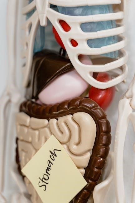
Digestive System: Enzyme Activity & Peristalsis
Lab manuals dedicated to human anatomy and physiology incorporate experiments demonstrating enzyme activity within the digestive system. These labs commonly investigate the breakdown of substrates – like starch or proteins – by specific enzymes such as amylase or pepsin, observing reaction rates under varying conditions (pH, temperature).
Students often utilize test tubes and spectrophotometry to quantify the products of enzymatic reactions, gaining a practical understanding of enzyme specificity and factors influencing their efficiency. Furthermore, labs explore peristalsis, the rhythmic contractions moving food through the digestive tract.
This is often modeled using simple tube systems and marbles, visually demonstrating the coordinated muscle contractions. The manual provides detailed protocols for these simulations, emphasizing the role of smooth muscle and the autonomic nervous system. Data analysis involves observing and recording the speed and effectiveness of peristaltic movement.
Ultimately, these experiments solidify understanding of digestive processes and enzyme function.
Urinary System: Urine Analysis
Human anatomy and physiology lab manuals feature comprehensive exercises centered around urine analysis, a crucial diagnostic tool. These labs guide students through both macroscopic and microscopic examination of urine samples, identifying key indicators of health and disease.
Typically, students assess urine color, clarity, odor, and pH using reagent strips, correlating these observations with potential physiological states. Microscopic analysis involves examining urine sediment for components like red blood cells, white blood cells, epithelial cells, and crystals.
The manual provides detailed instructions on proper slide preparation and identification of these elements under a microscope. Students learn to interpret findings in relation to kidney function, hydration levels, and potential infections. Data recording includes documenting all observations and drawing conclusions based on established norms.
These practical exercises reinforce understanding of renal physiology and the diagnostic significance of urinalysis.
Endocrine System: Hormone Level Analysis (Simulated)
Human anatomy and physiology lab manuals often incorporate simulated hormone level analysis to demonstrate the complexities of the endocrine system. Since direct hormone measurement is often beyond the scope of introductory labs, these exercises utilize case studies and data sets.
Students analyze simulated patient data, including symptoms and hormone concentrations (e.g., thyroid hormones, cortisol, insulin), to diagnose endocrine disorders. The manual provides background information on hormone functions and feedback loops, guiding students through the diagnostic process.
Exercises may involve interpreting graphs, calculating hormone ratios, and predicting the effects of hormone imbalances. Emphasis is placed on understanding the interplay between different endocrine glands and their impact on body functions.
These simulations enhance critical thinking skills and provide a foundational understanding of endocrine regulation, preparing students for more advanced studies.

Using Fetal Pig as a Model
Fetal pigs serve as excellent anatomical models, closely resembling human anatomy, making dissection valuable for human anatomy and physiology students.
This approach offers practical experience and a deeper understanding of organ systems.
Advantages of Fetal Pig Dissection
Fetal pig dissection presents numerous pedagogical advantages within a human anatomy and physiology laboratory setting. Their anatomical structures exhibit significant similarities to those of humans, particularly concerning organ systems like the circulatory, respiratory, and digestive systems.
This resemblance allows students to readily apply their knowledge of human anatomy to a tangible model, fostering a deeper comprehension of anatomical relationships and physiological functions. Furthermore, fetal pigs are readily available and relatively inexpensive compared to other dissection specimens, making them a practical choice for educational institutions.
The size of the fetal pig also contributes to its suitability; it’s large enough to allow for detailed observation of internal structures, yet manageable for students to work with effectively. Dissection encourages hands-on learning, enhancing critical thinking, problem-solving skills, and spatial reasoning abilities – all vital for future healthcare professionals.
Ethical Considerations in Dissection
Dissection, while valuable for learning human anatomy and physiology, necessitates careful consideration of ethical implications. Respectful handling of the specimen is paramount; students must approach the process with reverence and acknowledge the biological origin of the material.
Institutions should clearly outline the source of the fetal pigs and ensure they are obtained ethically, typically as a byproduct of the food industry. Alternatives to traditional dissection, such as virtual dissection software, should be offered to students who have conscientious objections.
Open discussion regarding these ethical concerns fosters a sense of responsibility and promotes critical thinking about the use of animals in scientific education. Proper disposal procedures, adhering to institutional guidelines and environmental regulations, are also crucial. Acknowledging these aspects ensures a balanced and ethically sound learning experience.